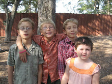
Jessie has been seizure free now for 1 1/2 months, and although we are overjoyed and feel very blessed, we are also grounded and realize that you never know what tomorrow will bring in the world of Epilepsy and our kids.
Nevertheless, I thought that I would share some information about the tissue that was removed and the symptoms that it resolved. I do this in hopes that other kids who may be dealing with the same types of recurring seizurs may benefit.
Jessie's returning seizures were characterized by:
1) Discomfort in the stomach (Aura if you will), followed by...
2) Partial, but not complete loss of cognitive awareness. She could still follow basic commands, nod yes or no. But she obviously wasn't "all there". She never fell to the ground.
3) Ipsilateral (same side as hemispherectomy) eye lid twitching. This was very scary, because we thought that the the seizures were coming from the contralateral good side of her brain. This just goes to show you that the "right controls the left and visa versa" isn't 100% for all tissue.
4) When it was over she heaved a heavy sigh, and wiped her nose.
5) Early on, the EEG was inconclusive. As the seizures progressed, the seizures were present, but they couldn't pinpoint 100% the origination point. Even with PET, SPECT, MRI, and EEG it was still a guess for the 2 Redo surgeries.
Jessie's original Hemispherectomy was anatomical. A word of caution that this doesn't necessarily mean everthing is taken out. It is not black and white, but more of a continuum when conceptualizing the varying degrees of hemispherectomy from "hemisphereotomy" to "Anatomical" and everything between, including "functional".
So the neurosurgeon at Cook Children's, in Jessie's first Re-do, tried to make sure everything was disconnected by retracing the corpus collosum. She was having seizures again soon after her first redo. It was obvious it didn't work. He didn't remove any tissue, because he felt that it was all disconnected.
The second Redo, the one that finally seems to have worked, went after small "button-sized" pieces of tissue in the frontal (Gyrys Rectus), Temporal, and Occipital areas. (I don't say lobes, because there were no lobes left from her original surgery).
The surgeon has decided, because of Jessie, that he plans to remove the Gyrus Rectus from now on in most hemispherectomy cases, especially Rasmussen's kids. This tissue, about the size of a few shirt buttons in the frontal lobe near midline seemed to be causing all of her problems.
Another reason that I want to share this, is that I was on Youtube last night, watching kids with post-hemi seizures, and some presented very similar to Jessie's. Her seizures are also on Youtube to compare. I know all kid's brains are different, but maybe this will help others, especially if the Gyrus Rectus does indeed tend to control ipsilateral eylid twitching.
See everyone in California for the Hemispherectomy Foundation Conference and Family Reunion!
Cris
Thursday, June 2, 2011
Subscribe to:
Post Comments (Atom)


3 comments:
Thank you, Cris, for posting this!!! You were on my "to-do" list this week to contact with a few questions because of similarities with my daughter's seizures. You answered the questions I had with this post and now I can ask my UCLA team to look into this. I know every case is different, but this is something that has been going on for years with my own daughter. Thank you Hall family for sharing your journey with us! Alexia Madigan
according to me,it's really a beautiful nd meaningfull site.atlanta rhinoplasty
i've been really behind on whats going on with Jessie. I am so happy for her and am trying to be as strong as you guys while we face a possible redo
Post a Comment